
Why Is sC5b-9 Difficult to Measure Accurately?
The complement system has three entrances but only one common terminal exit. The classical pathway, the lectin pathway, and the alternative pathway can be initiated by different triggers, but eventually converge on the C3 and C5 convertases, prompting the cleavage of C5 and initiating the sequential assembly of C5b, C6, C7, C8, and C9. Detection of soluble C5b-9 (sC5b-
The complement system has three entrances but only one common terminal exit. The classical pathway, the lectin pathway, and the alternative pathway can be initiated by different triggers, but eventually converge on the C3 and C5 convertases, prompting the cleavage of C5 and initiating the sequential assembly of C5b, C6, C7, C8, and C9. Detecting soluble C5b-9 (sC5b-9) is equivalent to observing whether the complement cascade has truly reached the terminal stage.
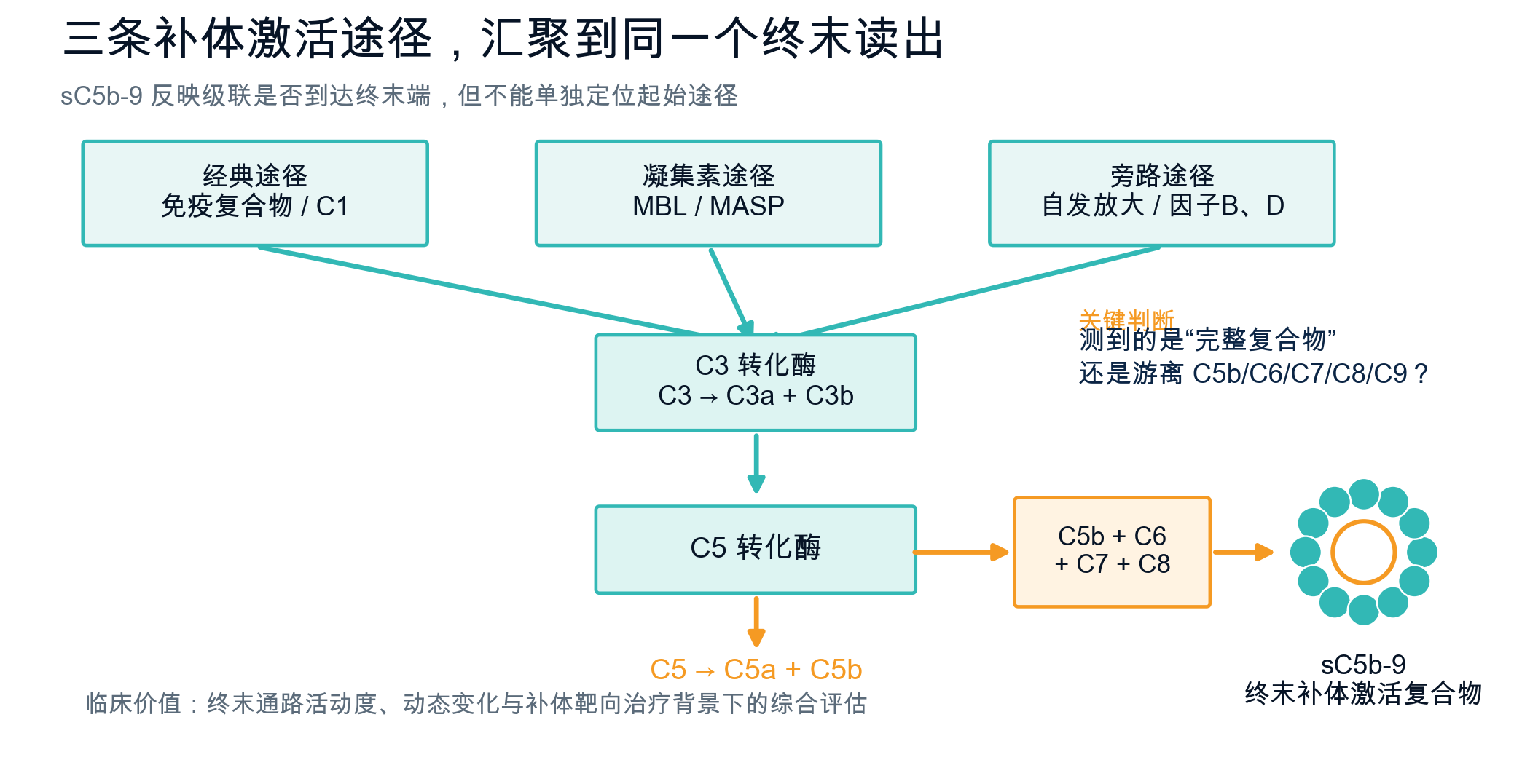
Figure 1 | Classical, lectin, and alternative pathways ultimately converge on C5 cleavage and C5b-9 assembly.
Three paths, one common destination
There are three entrances, why do we need a terminal indicator? Items such as C3, C4, and factor B can help describe complement reserves, consumption, or changes in a certain link, but the concentration of a single component does not mean that the pathway is being activated. sC5b-9 is a complex assembled from multiple terminal complement components: C5b-9 on the membrane can form pores, and the nascent complex in the liquid phase combines with regulatory molecules such as S protein/vitronectin and clusterin to form a soluble terminal complement complex. Thus, regardless of whether the initiating signal comes from the classical, lectin, or alternative pathway, as long as the cascade advances to the terminal end, sC5b-9 is likely to be formed. It can reflect "whether complement has reached the terminal effect stage", but it cannot clearly indicate "which pathway is activated first."
Clinically, what information does it provide?
Clinically, this “common endpoint” perspective is uniquely valuable. sC5b-9 is often used as an auxiliary indicator of terminal pathway activity in transplant-related thrombotic microangiopathy, atypical hemolytic-uremic syndrome, C3 glomerulopathy, and some transplant- and inflammation-related injuries. Studies have shown that sustained or early increases in some patients are related to disease activity, organ damage or adverse outcomes; in the context of complement-targeted therapy, continuous testing can also help to observe whether the terminal pathway is inhibited. However, sC5b-9 cannot yet be used as an independent diagnostic indicator and must be interpreted based on clinical manifestations, blood cell counts, LDH, renal function, urinary protein, C3/C4 and functional testing indicators.
Why is sC5b-9 so difficult to “pinpoint”?
The real difficulty is not to "measure C5, C6, C7, C8, and C9", but to confirm that what is measured is the assembled complex. There are inherently large amounts of free complement components in blood; if the capture or detection antibodies recognize a universal epitope on a monomeric protein, free C5b, C6, C7, C8 or C9 may interfere with the background signal. In vitro complement activation may also occur during sample collection, coagulation, transportation, and storage, further amplifying differences. Therefore, the comparability of results between different kits is affected by the combination of antibody epitopes, pairing strategies, calibrators, and pre-analytical procedures. The product name alone cannot determine whether it is truly specific for intact sC5b-9; one must look at evidence of cross-reactivity, dilution linearity, matrix effects, recovery, and sample stability.
Detect boundaries sC5b-9 can reflect terminal pathway activation but cannot independently differentiate between initiating sources of classical, lectin, or alternative pathways.
CompliTCC: Confirmation of the complete complex with two structural evidences
In the AlloDx CompliTCC sC5b-9 ELISA Kit, AlloDx uses a neo-epitope-specific double-antibody sandwich ELISA idea: one side of the antibody recognizes the conformational neo-epitope formed by polymerized C9, and the other side of the antibody recognizes the C5b domain. Only complete complexes with both "polymeric C9 signal" and "C5b domain" can form sandwich reads. The design goal is to exclude monomeric C9 and other free terminal complement components as much as possible, so that the test results are closer to the actual terminal complement activation state in the patient's body.
For clinical purposes, reliable sC5b-9 detection does not add one more test item, but provides a relatively direct evidence of "whether the three pathways have merged into the terminal pathway." Only by controlling the molecular state, antibody epitope and sample processing at the same time can sC5b-9 be transformed from a conceptual complement indicator into clinical information that can be used for dynamic assessment.
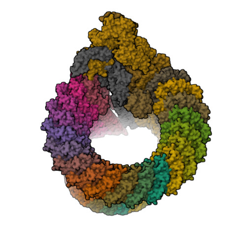
Figure 2 | The public PDB structure diagram is used to demonstrate the multi-component assembly characteristics of MAC/C5b-9; the diagram is not an analysis of the AD99 binding site.
References
1. Hugo F, Krämer S, Bhakdi S. Sensitive ELISA for quantitating terminal membrane C5b-9 and fluid-phase SC5b-9. J Immunol Methods. 1987;99:243-251. PMID: 3584995.
2. Chauvet S, et al. C3 and soluble C5b-9 levels and kidney outcomes in C3 glomerulopathy. Kidney Int. 2022;102:904-916. PMID: 35752323.
3. Horváth O, et al. Early increase in sC5b-9 predicts TA-TMA after stem cell transplantation. Biol Blood Marrow Transplant. 2018;24:989-996. PMID: 29339271.
4. Wilson RJ, et al. Validation and performance of MicroVue sC5b-9 Plus ELISA. Clin Chim Acta. 2025; PMID: 39788342.
Some original figures, videos and downloadable materials are provided in Chinese.