
Opportunities and challenges of Nephrin research (Part 1)
Recent studies have shown that anti-Nephrin autoantibodies (Anti-nephrin Ab) play an important role in the occurrence of minimal change disease (MCD) and focal segmental glomerulosclerosis (FSGS). This article outlines the blood circulation penetration of anti-Nephrin Ab as a component of MCD and primary FSGS (pFSGS).
Unexplained pFSGS
Recent studies have shown that anti-Nephrin autoantibodies (Anti-nephrin Ab) play an important role in the occurrence of minimal change disease (MCD) and focal segmental glomerulosclerosis (FSGS). This article outlines the research progress of anti-Nephrin antibodies as blood circulation penetration factors in MCD and primary FSGS (pFSGS), and provides prospects for future research directions.
The causes of MCD and FSGS are mainly divided into three categories: caused by genetic mutations in podocyte-related genes, secondary factors such as infection or drug use, and unexplained primary factors. Genetic mutations only account for about 8-14% [1-3], secondary factors account for 20-30% [4-5], and the rest are basically caused by unknown primary factors.Circulating Factor Theory and FSGS
When Hoyer et al. [6] first reported patients with recurrent FSGS after kidney transplantation in 1972, researchers speculated that certain circulating osmotic factors in the blood might cause the production of FSGS. Subsequent epidemiological studies have shown that FSGS after transplantation has a high recurrence rate, sometimes occurring within hours after transplantation. This result further prompted researchers to believe that the causative factor may not be a "local phenomenon within the kidney", but a blood circulation permeability factor that damages podocytes by changing the glomerular barrier [7-8]. Until 2012 Gallon et al[9]In a case study of a patient with recurrence of FSGS, it was found that when the transplanted kidney was removed and retransplanted to another recipient without a history of FSGS, the function of the kidney was restored, and the histopathological damage unique to FSGS was also reversed, which basically determined that certain circulating osmotic factors present in the blood caused the occurrence of FSGS.
In the following years, a large amount of research resources were devoted to finding circulating osmotic factors that play a key role in the formation of proteinuria in MCD and pFSGS [10-11]. Despite the investment of very large resources and research efforts, the identification of these unknown circulating permeability factors has been arduous and lengthy, and researchers have not found reliable and consistent biomarkers.
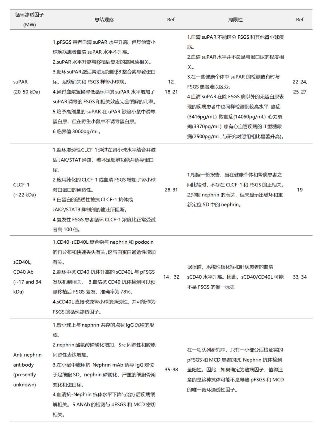
Some candidate biomarker molecules thought to increase serum protein vascular permeability (Table 1) include soluble urokinase-type plasminogen activator receptor (suPAR) [12], cardiotrophin-like cytokine-1 (CLCF-1) [13], soluble CD40 ligand [14], etc., have been found to be associated with the progression and recurrence of MCD and pFSGS, but studies to verify their clinical utility have shown that these markers do not play a decisive role in the development of MCD and FSGS, and they can also be detected in the serum of healthy and non-nephrotic syndrome patients [15-17].
table1. FSGS and MCD related circulating permeability factors and their limitations
Nephrin protein introduction
In 1966, in a group of Finnish families with congenital nephrotic syndrome (NPHS1) [39], it was discovered that this is a kidney disease caused by a rare genetic mutation, but the specific gene causing this disease is unknown. It was not until 1998 that the key chromosomal region where the NPHS1 gene was located was identified and sequenced. The expression product of the NPHS1 gene was named Nephrin, and its locus was located on chromosome 19q12-q13.1 [40-41]. Although the precise structure and function of the Nephrin protein remain unknown at this time, it is speculated that its domain structure is similar to a large class of cell adhesion receptors belonging to the immunoglobulin family, and that it can function as an adhesion receptor and signaling protein [40]. In subsequent studies [42-43], confirmed that Nephrin is a component of the podocyte slit diaphragm (SD)-related complex, which together with glomerular endothelial cells and basement membrane (GBM) forms the glomerular filtration barrier.(Figure 1)
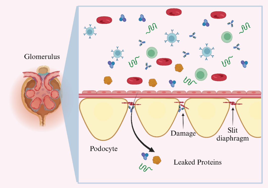
Figure 1. Nephrin antibodies induce mediastinal changes, podocyte damage, and protein leakage into urine.
Nephrin is a transmembrane protein with an extracellular fragment at its N-terminus and an intracellular domain at its C-terminus. The detection of an antibody specific for the N-terminus of human Nephrin confirmed that it is located in the mediastinum of the kidney [44]. The entire protein consists of eight immunoglobulin-like extracellular domains, a fibronectin type III-like domain, a transmembrane domain and a short intracellular domain. It maintains the tissue morphology, structure and function of podocytes through interaction with the actin cytoskeleton [40,45]. As a cell surface receptor protein of the immunoglobulin superfamily, Nephrin is also involved in intercellular adhesion and signaling functions. When Fyn of the Src kinase family phosphorylates one or more of the six tyrosine residues (Tyr1114, Tyr1136, Tyr1176, Tyr1183, Tyr1193, Tyr1217) of the intracellular domain of Nephrin, downstream signaling mediated by the intracellular tail of Nephrin is activated [46]. The specific performance is as followsThe phosphorylated intracellular domain interacts with several podocyte cytosolic proteins, including podocin, CD2AP, NEPH1, and phosphatidylinositol 3-kinase (PI3K), to transmit downstream signals to the actin cytoskeleton to regulate podocyte structural integrity and glomerular filtration.narrowThe function of sewing[47-48]. Phosphorylated Nephrin promotes the recruitment and interaction of Nck adapter proteins, ZO-1, catenin, barrel proteins, podocytes, CD2-AP, and PI3K. Nck adapter proteins send signals for actin remodeling and participate in the regulation of multiple intracellular signaling pathways, thereby affecting the polymerization dynamics of actin that is critical for podocyte stability [48-49] (Figure 2).
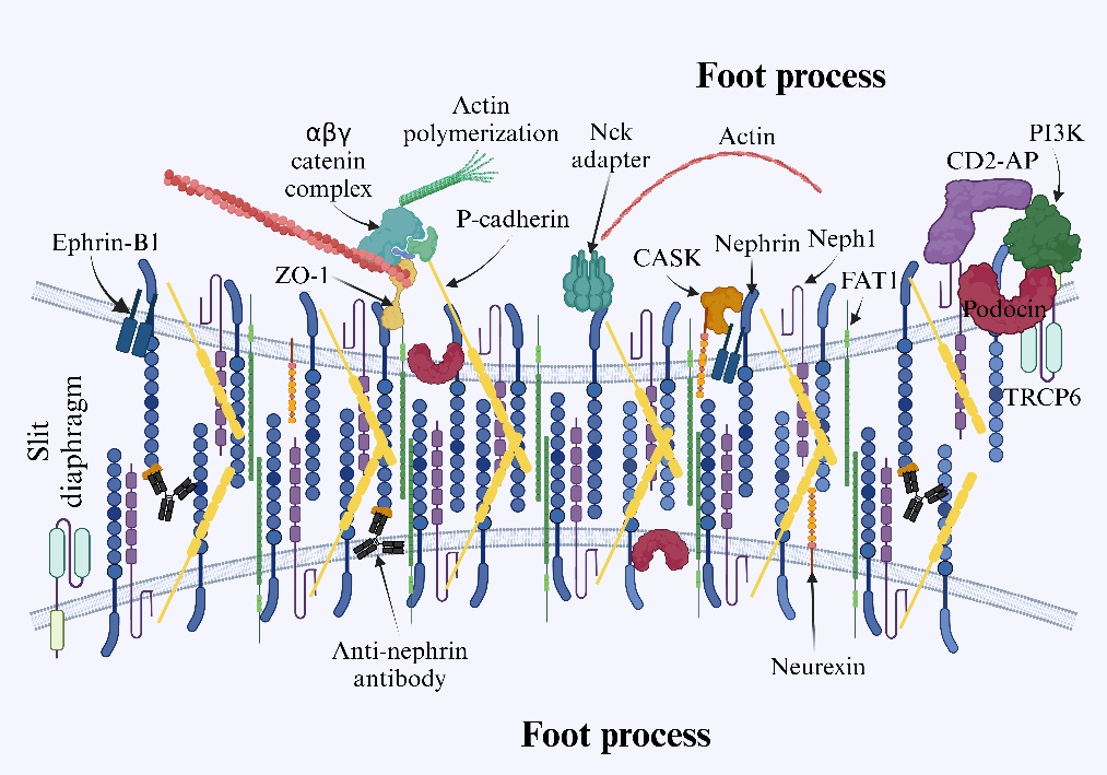
Figure 2. Schematic diagram of the gap layer between adjacent foot processes. The intracellular domain interacts with cytosolic proteins through cascade reactions when phosphorylated, inducing signaling, modulating actin polymerization dynamics and adjusting podocyte structure. The interaction between anti-Nephrin antibodies and Nephrin results in the loss of slit barrier function by causing Nephrin to relocate away from the slit layer.
References
[1]Büscher AK, Konrad M, Nagel M, et al. Mutations in podocyte genes are a rare cause of primary FSGS associated with ESRD in adult patients. Clin Nephrol. 2012;78(1):47-53. doi:10.5414/cn107320
[2]Isaranuwatchai S, Chanakul A, Ittiwut C, Ittiwut R, Srichomthong C, Shotelersuk V, Suphapeetiporn K, Praditpornsilpa K. Pathogenic variant detection rate by whole exome sequencing in Thai patients with biopsy-proven focal segmental glomerulosclerosis. Sci Rep. 2023 Jan 16;13(1):805. doi: 10.1038/s41598-022-26291-y. PMID: 36646731; PMCID: PMC9842604.
[3]Santín S, Bullich G, Tazón-Vega B, et al. Clinical utility of genetic testing in children and adults with steroid-resistant nephrotic syndrome.Clin J Am Soc Nephrol. 2011;6(5):1139-1148. doi:10.2215/CJN.05260610
[4]Gandzali Ngabe, P.E., Bonkano Baoua, D., Lengani, A.H.Y., Yattara, H., Kama Yatte, A., Loumingou, R., Tall, L., Ka, E.F., Niang, A. and Diouf, B. (2023) Focal Segmantal Glomerulosclerosis: Epidemiological, Clinico-Biological, Pathological, Etiological, Therapeutic and Evolutionary Profiles in Dakar. Open Journal of Nephrology, 13, 174-194. doi: 10.4236/ojneph.2023.132017.
[5]Rery TF Yuniarti, Ian Effendi, Zulkhair Ali, Novadian, Suprapti, Elfiani, Novandra AP, Dila Siti Hamidah, Fadil Pramudhya Husein, & Ika Kartika Edi P. (2024). Is It a Tumor or Not? A Case of Focal Segmental Glomerulosclerosis Secondary to Type 2 Diabetes with a Concomitant Renal Pseudotumor.Bioscientia Medicina : Journal of Biomedicine and Translational Research, 8(12), 5801-5813.
[6]Hoyer, J. R., Vernier, R. L., Najarian, J. S., Raij, L., Simmons, R. L., and Michael, A. F., Recurrence of idiopathic nephrotic syndrome after renal transplantation, Lancet, 1972, Vol 2(7773) p. 343-8.
[7]Dantal, J., Baatard, R., Hourmant, M., Cantarovich, D., Buzelin, F., and Soulillou, J. P., Recurrent nephrotic syndrome following renal transplantation in patients with focal glomerulosclerosis: a one-center study of plasma exchange effects, Transplantation, 1991, Vol 52(5) p.
[8]Savin Virginia, J., Sharma, R., Sharma, M., McCarthy Ellen, T., Swan Suzanne, K., Ellis, E., Lovell, H., Warady, B., Gunwar, S., Chonko Arnold, M., Artero, M., and Vincenti, F., Circulating Factor Associated with Increased Glomerular Permeability to Albumin in Recurrent Focal Segmental Glomerulosclerosis, New England Journal of Medicine, 1991, Vol 334(14) p. 878-883.
[9] Gallon, L., Leventhal, J., Skaro, A., Kanwar, Y., and Alvarado, A., Resolution of recurrent focal segmental glomerulosclerosis after retransplantation, N Engl J Med, 2012, Vol 366(17) p. 1648-9.
[10] den Braanker, D. J. W., Maas, R. J., Deegens, J. K., Yanginlar, C., Wetzels, J. F. M., van der Vlag, J., and Nijenhuis, T., Novel in vitro assays to detect circulating permeability factor(s) in idiopathic focal segmental glomerulosclerosis, Nephrology Dialysis Transplantation, 2021, Vol 36(2) p. 247-256.
[11] Gauckler, P., Shin, J. I., Alberici, F., Audard, V., Bruchfeld, A., Busch, M., Cheung, C. K., Crnogorac, M., Delbarba, E., Eller, K., Faguer, S., Galesic, K., Griffin, S., Hrušková, Z., Jeyabalan, A., Karras, A., King, C., Kohli, H. S., Maas, R., Mayer, G., Moiseev, S., Muto, M., Odler, B., Pepper, R. J., Quintana, L. F., Radhakrishnan, J., Ramachandran, R., Salama, A. D., Segelmark, M., Tesař, V., Wetzels, J., Willcocks, L., Windpessl, M., Zand, L., Zonozi, R., and Kronbichler, A., Rituximab in adult minimal change disease and focal segmental glomerulosclerosis - What is known and what is still unknown?, Autoimmunity Reviews, 2020, Vol 19(11) p. 102671.
[12] Wei, C., Trachtman, H., Li, J., Dong, C., Friedman, A. L., Gassman, J. J., McMahan, J. L., Radeva, M., Heil, K. M., Trautmann, A., Anarat, A., Emre, S., Ghiggeri, G. M., Ozaltin, F., Haffner, D., Gipson, D. S., Kaskel, F., Fischer, D. C., Schaefer, F., and Reiser, J., Circulating suPAR in two cohorts of primary FSGS, J Am Soc Nephrol, 2012, Vol 23(12) p. 2051-9.
[13] Sharma, M., Zhou, J., Gauchat, J. F., Sharma, R., McCarthy, E. T., Srivastava, T., and Savin, V. J., Janus kinase 2/signal transducer and activator of transcription 3 inhibitors attenuate the effect of cardiotrophin-like cytokine factor 1 and human focal segmental glomerulosclerosis serum on glomerular filtration barrier, Transl Res, 2015, Vol 166(4) p. 384-98.
[14] Doublier, S., Zennaro, C., Musante, L., Spatola, T., Candiano, G., Bruschi, M., Besso, L., Cedrino, M., Carraro, M., Ghiggeri, G. M., Camussi, G., and Lupia, E., Soluble CD40 ligand directly alters glomerular permeability and may act as a circulating permeability factor in FSGS, PLOS ONE, 2017, Vol 12(11) p. e0188045.
[15] Meijers, B., Maas, R. J. H., Sprangers, B., Claes, K., Poesen, R., Bammens, B., Naesens, M., Deegens, J. K. J., Dietrich, R., Storr, M., Wetzels, J. F. M., Evenepoel, P., and Kuypers, D., The soluble urokinase receptor is not a clinical marker for focal segmental glomerulosclerosis, Kidney International, 2014, Vol 85(3) p. 636-640.
[16] Müller-Deile, J., Sarau, G., Kotb, A. M., Jaremenko, C., Rolle-Kampczyk, U. E., Daniel, C., Kalkhof, S., Christiansen, S. H., and Schiffer, M., Novel diagnostic and therapeutic techniques reveal changed metabolic profiles in recurrent focal segmental glomerulosclerosis, Scientific Reports, 2021, Vol 11(1) p. 4577.
[17] Hou, S., Yang, B., Chen, Q., Xu, Y., and Li, H., Potential biomarkers of recurrent FSGS: a review, BMC Nephrology, 2024, Vol 25(1) p. 258.
[18]Wei, C., El Hindi, S., Li, J., Fornoni, A., Goes, N., Sageshima, J., Maiguel, D., Karumanchi, S. A., Yap, H. K., Saleem, M., Zhang, Q., Nikolic, B., Chaudhuri, A., Daftarian, P., Salido, E., Torres, A., Salifu, M., Sarwal, M. M., Schaefer, F., Morath, C., Schwenger, V., Zeier, M., Gupta, V., Roth, D., Rastaldi, M. P., Burke, G., Ruiz, P., and Reiser, J., Circulating urokinase receptor as a cause of focal segmental glomerulosclerosis, Nat Med, 2011, Vol 17(8) p. 952-60.
[19] Chebotareva, N., Vinogradov, A., Cao, V., Gindis, A., Berns, A., Alentov, I., and Sergeeva, N., Serum levels of plasminogen activator urokinase receptor and cardiotrophin-like cytokine factor 1 in patients with nephrotic syndrome, Clin Nephrol, 2022, Vol 97(2) p. 103-110.
[20] da Silva, C. A., Araújo, L. S., Dos Reis Monteiro, M. L. G., de Morais Pereira, L. H., da Silva, M. V., Castellano, L. R. C., Corrêa, R. R. M., Dos Reis, M. A., and Machado, J. R., Evaluation of the Diagnostic Potential of uPAR as a Biomarker in Renal Biopsies of Patients with FSGS, Dis Markers, 2019, Vol 2019 p. 1070495.
[21] Wei, C., Möller, C. C., Altintas, M. M., Li, J., Schwarz, K., Zacchigna, S., Xie, L., Henger, A., Schmid, H., Rastaldi, M. P., Cowan, P., Kretzler, M., Parrilla, R., Bendayan, M., Gupta, V., Nikolic, B., Kalluri, R., Carmeliet, P., Mundel, P., and Reiser, J., Modification of kidney barrier function by the urokinase receptor, Nat Med, 2008, Vol 14(1) p. 55-63. 10.1038/nm1696.
[22] Meijers, B., Maas, R. J. H., Sprangers, B., Claes, K., Poesen, R., Bammens, B., Naesens, M., Deegens, J. K. J., Dietrich, R., Storr, M., Wetzels, J. F. M., Evenepoel, P., and Kuypers, D., The soluble urokinase receptor is not a clinical marker for focal segmental glomerulosclerosis, Kidney International, 2014, Vol 85(3) p. 636-640.
[23] Maas, R. J. H., Wetzels, J. F. M., and Deegens, J. K. J., Serum-soluble urokinase receptor concentration in primary FSGS, Kidney International, 2012, Vol 81(10) p. 1043-1044.
[24] Maas, R. J. H., Wetzels, J. F. M., and Deegens, J. K. J., Serum suPAR concentrations in patients with focal segmental glomerulosclerosis with end-stage renal disease, Kidney International, 2014, Vol 85(3) p. 711.
[25] Maas, R. J. H., Deegens, J. K. J., and Wetzels, J. F. M., Serum suPAR in patients with FSGS: trash or treasure?, Pediatric Nephrology, 2013, Vol 28(7) p. 1041-1048. 10.1007/s00467-013-2452-5.
[26] Hayek, S. S., Tahhan, A. S., Ko, Y.-A., Alkhoder, A., Zheng, S., Bhimani, R., Hartsfield, J., Kim, J., Wilson, P., Shaw, L., Wei, C., Reiser, J., and Quyyumi, A. A., Soluble Urokinase Plasminogen Activator Receptor Levels and Outcomes in Patients with Heart Failure, Journal of Cardiac Failure, 2023, Vol 29(2) p. 158-167.
[27] Mohammed, M. S. and Ahmed, H. S., Plasminogen activator urokinase receptor as a diagnostic and prognostic biomarker in type 2 diabetic patients with cardiovascular disease, J Cardiovasc Thorac Res, 2023, Vol 15(3) p. 154-160. 10.34172/jcvtr.2023.32895.
[28] Sharma, M., Zhou, J., Gauchat, J. F., Sharma, R., McCarthy, E. T., Srivastava, T., and Savin, V. J., Janus kinase 2/signal transducer and activator of transcription 3 inhibitors attenuate the effect of cardiotrophin-like cytokine factor 1 and human focal segmental glomerulosclerosis serum on glomerular filtration barrier, Transl Res, 2015, Vol 166(4) p. 384-98.
[29] Savin, V. J., Sharma, M., Zhou, J., Gennochi, D., Fields, T., Sharma, R., McCarthy, E. T., Srivastava, T., Domen, J., Tormo, A., and Gauchat, J. F., Renal and Hematological Effects of CLCF-1, a B-Cell-Stimulating Cytokine of the IL-6 Family, J Immunol Res, 2015, Vol 2015 p. 714964.
[30] Savin, V. J., McCarthy, E. T., and Sharma, M., Permeability factors in nephrotic syndrome and focal segmental glomerulosclerosis, Kidney Res Clin Pract, 2012, Vol 31(4) p. 205-13.
[31] McCarthy, E. T., Sharma, M., and Savin, V. J., Circulating permeability factors in idiopathic nephrotic syndrome and focal segmental glomerulosclerosis, Clin J Am Soc Nephrol, 2010, Vol 5(11) p. 2115-21.
[32] Delville, M., Sigdel, T. K., Wei, C., Li, J., Hsieh, S. C., Fornoni, A., Burke, G. W., Bruneval, P., Naesens, M., Jackson, A., Alachkar, N., Canaud, G., Legendre, C., Anglicheau, D., Reiser, J., and Sarwal, M. M., A circulating antibody panel for pretransplant prediction of FSGS recurrence after kidney transplantation, Sci Transl Med, 2014, Vol 6(256) p. 256ra136.
[33] Komura, K., Fujimoto, M., Matsushita, T., Yanaba, K., Kodera, M., Kawasuji, A., Hasegawa, M., Takehara, K., and Sato, S., Increased serum soluble CD40 levels in patients with systemic sclerosis, J Rheumatol, 2007, Vol 34(2) p. 353-8.
[34] Liu, H., Qi, C. J., Zhuang, Y. M., Gan, J. H., Li, H. L., Yin, C. S., and Zhang, X. G., [Serum levels and clinical significance of soluble CD40 in liver disease], Xi Bao Yu Fen Zi Mian Yi Xue Za Zhi, 2006, Vol 22(6) p. 738-41.
[35] Chebotareva, N., Vinogradov, A., Birukova, Y., Alentov, I., Sergeeva, N., Chemodanova, D., Kononikhin, A. S., and Moiseev, S. V., A pilot study of anti-nephrin antibodies in podocytopaties among adults, Nephrology (Carlton), 2024, Vol 29(2) p. 86-92.
[36] Hengel Felicitas, E., Dehde, S., Lassé, M., Zahner, G., Seifert, L., Schnarre, A., Kretz, O., Demir, F., Pinnschmidt Hans, O., Grahammer, F., Lucas, R., Mehner Lea, M., Zimmermann, T., Billing Anja, M., Oh, J., Mitrotti, A., Pontrelli, P., Debiec, H., Dossier, C., Colucci, M., Emma, F., Smoyer William, E., Weins, A., Schaefer, F., Alachkar, N., Diemert, A., Hogan, J., Hoxha, E., Wiech, T., Rinschen Markus, M., Ronco, P., Vivarelli, M., Gesualdo, L., Tomas Nicola, M., and Huber Tobias, B., Autoantibodies Targeting Nephrin in Podocytopathies, New England Journal of Medicine, 2024, Vol 391(5) p. 422-433.
[37] Watts, A. J. B., Keller, K. H., Lerner, G., Rosales, I., Collins, A. B., Sekulic, M., Waikar, S. S., Chandraker, A., Riella, L. V., Alexander, M. P., Troost, J. P., Chen, J., Fermin, D., Yee, J. L., Sampson, M. G., Beck, L. H., Jr., Henderson, J. M., Greka, A., Rennke, H. G., and Weins, A., Discovery of Autoantibodies Targeting Nephrin in Minimal Change Disease Supports a Novel Autoimmune Etiology, J Am Soc Nephrol, 2022, Vol 33(1) p. 238-252.
[38] Shirai, Y., Miura, K., Ishizuka, K., Ando, T., Kanda, S., Hashimoto, J., Hamasaki, Y., Hotta, K., Ito, N., Honda, K., Tanabe, K., Takano, T., and Hattori, M., A multi-institutional study found a possible role of anti-nephrin antibodies in post-transplant focal segmental glomerulosclerosis recurrence, Kidney Int, 2024, Vol 105(3) p. 608-617.
[39] Norio, R., Heredity in the congenital nephrotic syndrome. A genetic study of 57 finnish FAMILIES WITH A REVIEW OF REPORTED CASES, Ann Paediatr Fenn, 1966, Vol 12 p. Suppl 27:1-94.
[40] Kestilä, M., Lenkkeri, U., Männikkö, M., Lamerdin, J., McCready, P., Putaala, H., Ruotsalainen, V., Morita, T., Nissinen, M., Herva, R., Kashtan, C. E., Peltonen, L., Holmberg, C., Olsen, A., and Tryggvason, K., Positionally Cloned Gene for a Novel Glomerular Protein—Nephrin—Is Mutated in Congenital Nephrotic Syndrome, Molecular Cell, 1998, Vol 1(4) p. 575-582.
[41] Kestilä, M., Männikkö, M., Holmberg, C., Gyapay, G., Weissenbach, J., Savolainen, E. R., Peltonen, L., and Tryggvason, K., Congenital nephrotic syndrome of the Finnish type maps to the long arm of chromosome 19, Am J Hum Genet, 1994, Vol 54(5) p. 757-64.
[42] Ruotsalainen, V., Ljungberg, P., Wartiovaara, J., Lenkkeri, U., Kestilä, M., Jalanko, H., Holmberg, C., and Tryggvason, K., Nephrin is specifically located at the slit diaphragm of glomerular podocytes, Proc Natl Acad Sci U S A, 1999, Vol 96(14) p. 7962-7.
[43]Topham, P. S., Kawachi, H., Haydar, S. A., Chugh, S., Addona, T. A., Charron, K. B., Holzman, L. B., Shia, M., Shimizu, F., and Salant, D. J., Nephritogenic mAb 5-1-6 is directed at the extracellular domain of rat nephrin, J Clin Invest, 1999, Vol 104(11) p. 1559-66.
[44Holzman, L. B., St John, P. L., Kovari, I. A., Verma, R., Holthofer, H., and Abrahamson, D. R., Nephrin localizes to the slit pore of the glomerular epithelial cell, Kidney Int, 1999, Vol 56(4) p. 1481-91.
[45] Schoeb, D. S., Chernin, G., Heeringa, S. F., Matejas, V., Held, S., Vega-Warner, V., Bockenhauer, D., Vlangos, C. N., Moorani, K. N., Neuhaus, T. J., Kari, J. A., MacDonald, J., Saisawat, P., Ashraf, S., Ovunc, B., Zenker, M., and Hildebrandt, F., Nineteen novel NPHS1 mutations in a worldwide cohort of patients with congenital nephrotic syndrome (CNS), Nephrol Dial Transplant, 2010, Vol 25(9) p. 2970-6.
[46] Denhez, B. and Geraldes, P., "Regulation of Nephrin Phosphorylation in Diabetes and Chronic Kidney Injury", in Protein Reviews: Volume 18, M.Z. Atassi, Editor, Springer Singapore: Singapore. 2017. p. 149-161.
[47] Tian, Y., Chen, X.-m., Liang, X.-m., Wu, X.-b., and Yao, C.-m., SGLT2 inhibitors attenuate nephrin loss and enhance TGF-β1 secretion in type 2 diabetes patients with albuminuria: a randomized clinical trial, Scientific Reports, 2022, Vol 12(1) p. 15695.
[48] Verma, R., Kovari, I., Soofi, A., Nihalani, D., Patrie, K., and Holzman, L. B., Nephrin ectodomain engagement results in Src kinase activation, nephrin phosphorylation, Nck recruitment, and actin polymerization, J Clin Invest, 2006, Vol 116(5) p. 1346-59.
[49] Jones, N., Blasutig, I. M., Eremina, V., Ruston, J. M., Bladt, F., Li, H., Huang, H., Larose, L., Li, S. S. C., Takano, T., Quaggin, S. E., and Pawson, T., Nck adaptor proteins link nephrin to the actin cytoskeleton of kidney podocytes, Nature, 2006, Vol 440(7085) p. 818-823.
Some original figures, videos and downloadable materials are provided in Chinese.