
Growing Interest in Nephrin Antibodies: Choosing a Testing Method
Nephrin antibodies, as a new circulating penetrant factor in podocyte nephropathy, are considered to be of great value in distinguishing MCD (minimal change kidney disease)/FSGS (focal segmental glomerulosclerosis) related to immune factors from non-immune factors. However, there are currently many challenges in the detection and quantification of Nephrin antibodies. human serum pack
Nephrin antibodies, as a new circulating penetrant factor in podocyte nephropathy, are considered to be of great value in distinguishing MCD (minimal change kidney disease)/FSGS (focal segmental glomerulosclerosis) related to immune factors from non-immune factors. However, there are currently many challenges in the detection and quantification of Nephrin antibodies. Human serum contains tens of thousands of proteins and antibodies, and the Nephrin antibody content is extremely low and is easily interfered by high-abundance proteins and easily combined with other impurities in the serum, leading to false negative results.
Various international Nephrin detection teams have reported a variety of strategies for detecting Nephrin antibodies, including immunoprecipitation using recombinant human glomerular basement membrane (Figure 1A), combining signal-enhanced ELISA with recombinant human glomerular basement membrane (Figure 1B, left), using traditional ELISA, and a combined method of immunoprecipitation combined with ELISA to quantify the precipitated recombinant glomerular basement membrane (Figure 1B, right) etc.
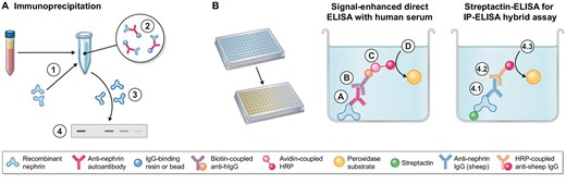
Figure 1:Detection of Nephrin antibodies.
(A) Overview of immunoprecipitation for Nephrin antibody detection in human serum samples. (1) Human serum is incubated with recombinant glomerular basement membrane protein to form antibody-antigen complexes. (2) Add antibody-binding resin or beads to precipitate antibody-antigen complexes. (3) The precipitated protein is eluted to release the antibody and bound antigen. (4) Gel electrophoresis and western blotting can be used to specifically detect precipitated glomerular basement membrane proteins in samples in which recombinant proteins have been precipitated.
(B) Overview of different ELISA techniques for the detection of Nephrin antibodies in human serum samples.(left)Signal-enhanced direct ELISA uses immobilized recombinant glomerular basement membrane to which human antibodies are bound (A) and detected by biotin-linked anti-human immunoglobulin G (IgG) antibodies (B). It is then bound via avidin-linked horseradish peroxidase (C). Horseradish peroxidase catalyzes the conversion of a chromogenic substrate, indicating the amount of bound human antibody (D).(right)Immunoprecipitation is followed by hybridization detection by ELISA via binding of its avidin tag to the streptactin-ELISA plate and specific detection of bound glomerular basement membrane using a commercial sheep-derived Nephrin antibody (4.1) followed by binding to a horseradish peroxidase-conjugated secondary anti-sheep IgG antibody (4.2), which is quantified by a chromogenic substrate (4.3).
Recently, the German Hengel team published a review in the journal CKJ, providing an in-depth analysis of the impact of different Nephrin antibody detection methods on clinical research results (Table 1).
Table 1:Overview of detection methods and detection rates of circulating Nephrin antibodies in different studies and cohorts
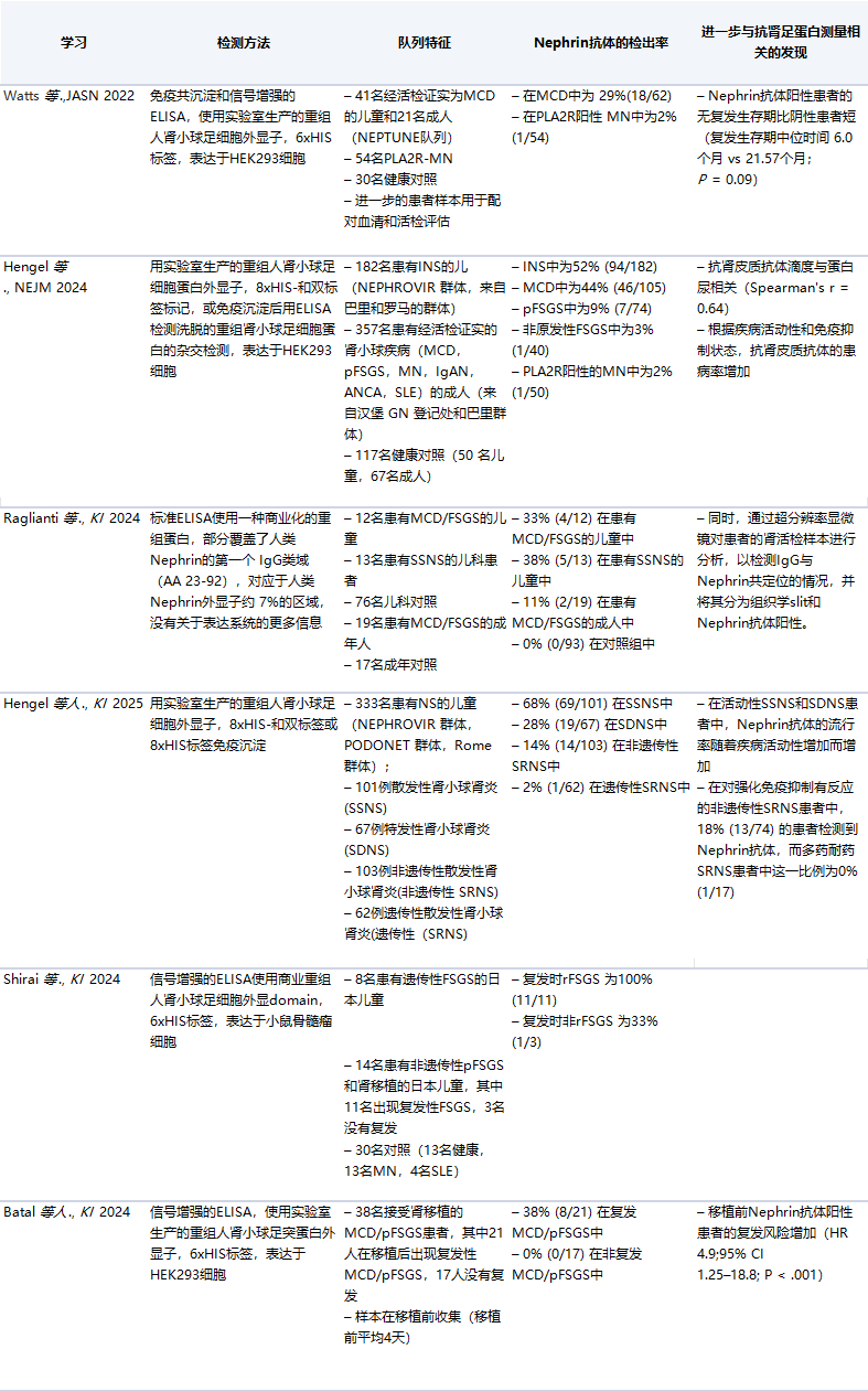
JASN, Journal of the American Society of Nephrology; NEJM, New England Journal of Medicine; KI, Kidney International; HEK293, human embryonic kidney cell 293; MN, membranous nephropathy; pFSGS, primary FSGS; IgAN, IgA nephropathy; ANCA, antineutrophil cytoplasmic antibodies; SLE, systemic lupus erythematosus; SSNS, steroid-sensitive nephrotic syndrome; SDNS, steroid-dependent nephrotic syndrome; HR, hazard ratio; CI, confidence interval.
Hengel's team also conducted a detailed comparative analysis of the advantages and disadvantages of currently reported Nephrin detection methods:
1.Co-immunoprecipitation(Co-IP)
Compared with traditional antibody detection methods, it shows significant advantages:(1) high sensitivity, which can effectively detect low-level circulating autoantibodies through antibody-binding resin/bead enrichment and purification of patient antibodies;(2) high specificity, a positive result requires the formation of two independent antibody-antigen complexes (the serum autoantibody binds to the target antigen in the solution, and the target antigen-specific antibody detects the precipitated antigen). Although in theory, the antigen-binding resin may be risky, it can be controlled through appropriate procedures, and combined with Western Blot detection, the antigen molecular weight information can be further used to improve specificity and reduce false positives;(3) can detect weak interactions, forming an antigen-autoantibody complex under natural conditions (without denaturants/reducing agents or antigen fixation), avoiding the reduction or loss of epitope binding caused by processes such as Western sample preparation or ELISA. These characteristics make Co-IP excellent at detecting autoantibodies, especially low-level antibodies. However, it also has some limitations, such as high sample requirements (e.g. 30µL/detection), cumbersome and time-consuming operation, and only semi-quantification through Western Blot.
2.Standard ELISA
Standard ELISA (such as using recombinant glomerular factor coating) has the advantages of simple operation, high throughput, low sample requirements (usually 1-2µL/assay), and the ability to quantify antibodies through a standard curve. However, multiple studies have shown that standard ELISA cannot reliably quantify Nephrin antibodies, possibly because the Nephrin antibody content is too low and requires an additional signal amplification step. Signal amplification may increase non-specific signals, leading to false positive results.
Three.Co-immunoprecipitation combined with ELISA
Co-immunoprecipitation combined with ELISA can overcome the shortcomings of the two methods to a certain extent by quantifying the precipitated recombinant Nephrin (Figure 1B, right), achieving sensitive and specific quantitative measurement of Nephrin titer. However, this method still requires a lot of manual work and has higher sample size requirements than the standard ELISA method.
4. Other information about Nephrin antibodiesDetection method
Other detection methods for Nephrin antibodies also have many shortcomings: first, the use of commercial recombinant glomerular podocin or different protein tags, which are different from human-expressed glomerular podocin, or lack of information on the expression system, leading to high false positive results; second, co-staining of IgG and glomerular basement membrane on frozen kidney biopsy specimens and co-localization. This technology only suggests a possible antigen through co-localization and does not clearly indicate that the target antigen of IgG is the glomerular basement membrane. The third is cell-based fluorescence technology. Although it can simulate the conditions for Nephrin antibodies to bind near the cell membrane, it is still quite different from the local conditions at the native pore of the glomerular filtration barrier.
Taken together, Nephrin antibody detection methods based on antibody capture and enrichment pathways can improve the stability and reproducibility of Nephrin antibody detection, thereby bringing potential benefits in diagnosis, prognosis and treatment guidance.The Super Nephrin Antibody Trap (SuperNAT) second-generation highly sensitive Nephrin antibody detection solution developed by domestic AlloDx, based on the antibody enrichment platform of SuperNAT technology, significantly reduces the background noise of Nephrin antibody detection and increases the detection rate by 53% compared with the traditional ELISA method.
Some original figures, videos and downloadable materials are provided in Chinese.