
2022ATC in-depth interpretation (3): Factors affecting ddcfDNA detection values
As ddcfDNA enters the clinical application stage of transplantation, it is necessary to correctly interpret the clinical significance of the detection values. On the one hand, we are required to understand the change pattern characteristics of ddcfDNA in different pathological states. On the other hand, we also need to understand the impact of non-pathological factors on the detection values of ddcfDNA. There are 13 representative research papers at this ATC conference.
As ddcfDNA enters the clinical application stage of transplantation, it is necessary to correctly interpret the clinical significance of the detection values. On the one hand, we are required to understand the change pattern characteristics of ddcfDNA in different pathological states. On the other hand, we also need to understand the impact of non-pathological factors on the detection values of ddcfDNA. There are 13 representative studies at this ATC conference focusing on the theme of "factors affecting ddcfDNA detection values". Next, we will summarize it into three parts for detailed interpretation.
What impact do donor/recipient demographic characteristics, different donor types, and preoperative induction regimens have on early postoperative ddcfDNA values?
Augusta University Medical Center, Augusta, GA found that combined multi-organ transplantation and male donors resulted in higher ddcfDNA values 1 month after transplantation. Through the detection of ddcfDNA in 101 kidney transplant patients 1 month after surgery, it was found that the median ddcfDNA value of kidney-pancreas dual transplant was significantly higher than that of single kidney transplant (1.5% vs 0.4%, n=3 vs 98, p < 0.05) (Figure 1a). The median recipient ddcfDNA value of male donors was significantly higher than that of female donors (0.575% vs 0.28%, n = 62 vs 38, p < 0.01) (Fig. 1b).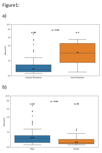
(Figure 1)
In heart transplantation, research by Professor A. Alam’s team at Baylor University Medical Center, Dallas, TX, found that the gender/race factors of the donor/recipient did not have a significant impact on the ddcfDNA value 3 months after surgery (Figure 2). The study enrolled 609 heart transplant patients who were stable within one year after surgery (not all donor/recipient characteristics were included in the analysis), of which 189 had ddcfDNA test results within 3 months after surgery, with a median value of 0.053% (IQR: 0.028-0.098).
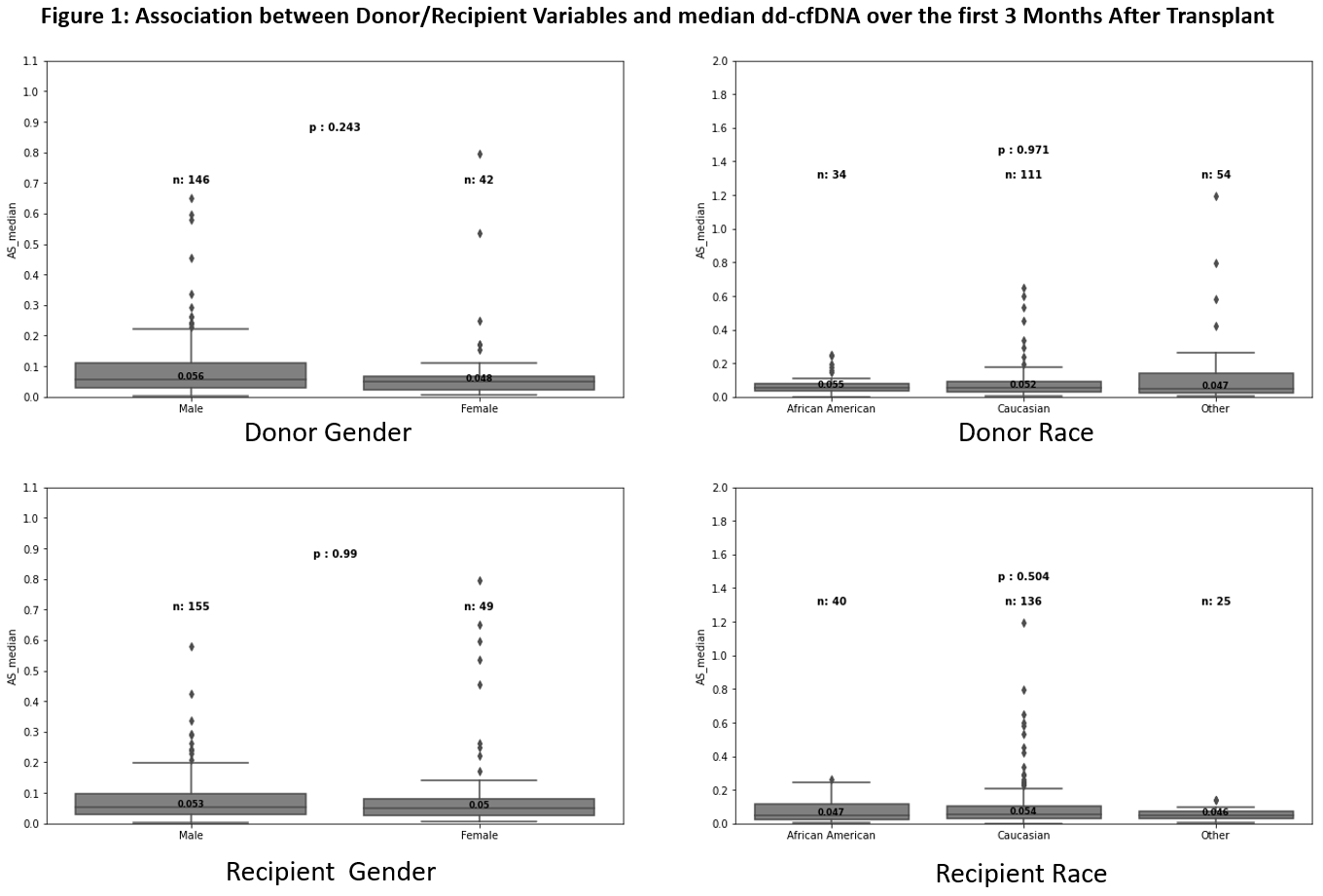
(Figure 2)
When analyzing the age factor, no significant correlation was found between the ddcfDNA value and recipient age. Even in the older recipient group (51-70 years old), the ddcfDNA value was still relatively low (0.047%, IQR: 0.029-0.083). However, there was a weak correlation between ddcfDNA value and donor age, and it was statistically significant (Pearson's r: 0.178, p = 0.014) (Figure 3).
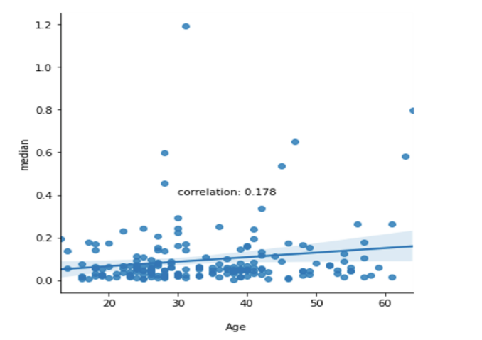
(Figure 3)
In the multifactor (sex, age and race) model, no synergistic factors were found to significantly affect the ddcfDNA value 3 months after surgery, and the overall predictive ability was poor (R2 = 0.059).
This study concluded that donor/recipient demographic characteristics (gender, age, and race) were not significantly related to ddcfDNA values 3 months after heart transplantation, suggesting that the significant increase in ddcfDNA in the early post-transplantation period could be interpreted as caused by specific immune damage.
Similarly, research data from Columbia University Irving Medical Center, New York also shows that gender has no significant impact on ddcfDNA values in heart transplant recipients, and ddcfDNA test values do not need to be interpreted specifically for gender.
A total of 2029 recipients (544 women, 26.8%) were enrolled in the study, 67.7% of whom were white, with an average age of 56.7 years. 8.5% of recipients were hepatitis C-positive donors, and 58.1% of women and 53.5% of men were infected with CMV. 56.1% of female recipients had female donors, while only 24.5% of male recipients had female donors.
There was no significant difference in the median ddcfDNA values between male and female recipients at steady state (p=0.133) (Figure 4). There were 132 (23.7% female) recipients who experienced rejection (49 cases of AMR, 77 cases of ACR, and 6 cases of ACR combined with AMR). In the 90 days before rejection was diagnosed, there was no significant difference in ddcfDNA values among different genders (106 samples) (Figure 5).
At a median follow-up of 357 days, there were no significant differences in ddcfDNA values between sexes for outcomes affected by rejection, dnDSA, CAV, or decreased LVEF (85 women, 17.7%; 260 men, 15.9%; log-rank p = 0.36).
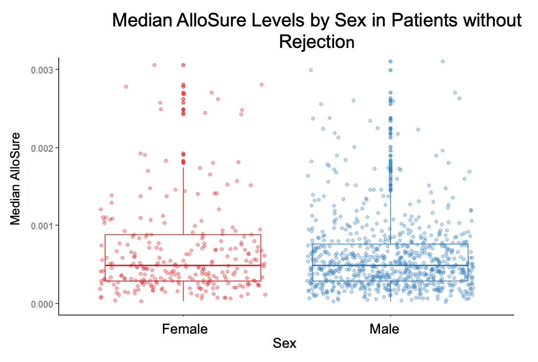
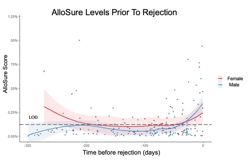 (Figure 5)
Regarding the impact of racial differences on ddcfDNA detection values, some studies have shown that in the stable state after transplantation, the ddcfDNA values of African American (AA) recipients are higher than those of non-African Americans (non-AA). Researchers from Rush University Medical Center, Chicago, IL, further analyzed the difference in ddcfDNA values between AA and non-AA recipients when rejection occurred.
The study enrolled 40 kidney transplant recipients (19 AA, 21 non-AA) with a total of 55 biopsy results (27 AA, 28 non-AA). 28 had rejection (15 AA, 13 non-AA) and 27 had no rejection (12 AA, 15 non-AA). The median value of ddcfDNA in the rejection group was 1.45% (IQR 0.53%-2.5%), the median value in the AA rejection group was 1.30% (IQR 0.38-1.8%), and the median value in the non-AA rejection group was 2.3% (IQR 0.89-4.4%). The median value of ddcfDNA in the non-rejection group was 0.24% (IQR 0.15-0.47%), and the median value in the AA non-rejection group was 0.2% (IQR 0.15-0.67%) (Figure 6). The area under the curve (AUC) of the rejection receiver operating curve (ROC) analysis for the entire cohort was 0.84 (95% CI = 0.74-0.95), the AUC for the AA alone group was 0.74 (95% CI = 0.54-0.95), and the AUC for the non-AA group was 0.95 (95% CI = 0.88-1.00) (Figure 7).
It is worth noting that among recipients who experienced rejection, 33% (n = 5: 3 ACR, 1 AMR, 1 mixed) of AA recipients had ddcfDNA <0.5%, while only 0.08% (n = 1, AMR) of non-AA recipients had ddcfDNA <0.5%. This suggests that when AA recipients and non-AA recipients have the same low levels of ddcfDNA, rejection may have occurred in AA recipients. The reliability of this difference requires further study.
(Figure 6)(Figure 7)
In addition to studies on donor/recipient demographic characteristics, there are also research reports on the effects of different donor types, cold ischemia time (CIT), and different immune induction protocols on early postoperative ddcfDNA detection values.
The Department of Nephrology, Medicine Institute, Allegheny General Hospital, Pittsburgh conducted a retrospective study on 111 kidney transplant recipients (including recipients with ddcfDNA <1.0%) who underwent ddcfDNA testing approximately 8 weeks after transplantation. Among them, 39 cases were donated by LDRT, 55 cases by DBD, and 17 cases by DCD; 63 cases had CIT <12 hours, 46 cases had CIT ≥12 hours, and 2 cases had missing CIT data; 101 cases used ATG, and 10 cases used basiliximab. The study found that there was no significant difference in ddcfDNA detection values among different donor types, CIT times, and different immune induction protocols (Figure 8).
(Figure 8)
Early postoperative ischemia-reperfusion injury (IRI), including acute tubular injury/necrosis (ATI/ATN) caused by it, is a common non-rejection type of injury after surgery. Studies have shown that IRI can cause an increase in ddcfDNA values. Is there any difference in its increasing trend in different types of recipients? There are 5 studies in this ATC focusing on this direction.
Studies have shown that ddcfDNA in adult kidney transplant recipients shows an L-shaped downward trend after surgery. A study from Pediatrics, Washington University School of Medicine in St. Louis, St. Louis, MO, explored the changes in ddcfDNA detection values in pediatric kidney transplant recipients in the early postoperative period. The results showed that the kinetics of postoperative ddcfDNA values in pediatric kidney transplant recipients were different from those in adults. The ddcfDNA values in pediatric recipients remained at a high level for a long time after surgery.
Through dynamic monitoring of ddcfDNA in 71 pediatric recipients (30 days to 1 year after surgery), 204 samples from 54 stable recipients were screened, and samples such as major infections, rejection reactions, and delayed kidney transplant function were excluded.
In this cohort, the median ddcfDNA value at 30 days after transplantation was 0.84 (0.64-1.2)%, decreasing to 0.43 (0.25-0.66)% by 2 months and 0.23 (0.16-0.32)% by 6 months. When further grouped according to recipient age (>10 years, <10 years), the mean ddcfDNA value in the <10 years group (67 samples) was significantly higher in the first year after transplantation compared with the >10 years group (137 samples). The median ddcfDNA value in the <10-year-old group fell below 0.2% 12 months after transplantation, while the median ddcfDNA value in the >10-year-old group dropped below 0.2% within 4 months after transplantation (Figure 9).
(Figure 9)
Research from the Medical City Transplant Institute, Fort Worth suggests that acute tubular injury/necrosis in the early postoperative period will not affect the diagnosis of rejection using ddcfDNA values. Using ddcfDNA values to identify non-rejection injuries (including ATI/ATN) can help reduce the number of unnecessary biopsies.
The study screened samples with paired ddcfDNA results within 30 days of needle biopsy and divided them into a stable group (141 patients, 166 biopsy results) and an ATI/ATN group (64 patients, 70 biopsy results).
Compared with the ATI/ATN group, the stable group had a lower median KDPI (IQR 49% vs 64%, p < 0.05) and shorter cold ischemia time (13 vs 18 hours, p < 0.01). The ATI/ATN group underwent biopsy earlier (83.0 days vs 116.5 days, p<0.001) and had lower eGFRs (32 vs 43mL/min, p<0.001) (Figure 10). There was no significant difference in the median ddcfDNA values between the stable group and the ATI/ATN group (0.23%, IQR: 0.11-0.53 vs 0.21%, IQR: 0.13-0.55, p = 0.993) (Figure 11). Further grouping of ddcfDNA values during the first biopsy (<0.5% vs ≥0.5%) found that there was no significant difference in the incidence of complications 1 year after surgery between the two groups (12.9% vs 27.5%, p=0.53), while eGFR decline was the most common in the ≥0.5% group (78.5% incidence).
(Figure 10)(Figure 11)
In a study of ischemia-reperfusion injury after liver transplantation, results from Mount Sinai Medical Center, New York, found that ddcfDNA values decreased exponentially in liver transplant recipients after surgery, and this downward trend was faster in living donor liver transplant recipients. Fourteen days after liver transplantation, the recipient's ddcfDNA value can return to a stable low level without rejection, graft dysfunction, or infection.
This study enrolled 31 stable liver transplant recipients and conducted 114 tests within 6 months after surgery. It was found that the ddcfDNA value increased significantly in the first week after surgery (median 22.3%, IQR 12.91-33.38%) and declined rapidly within 2 weeks (Figure 12A). In living donor liver transplant recipients, the median ddcfDNA value decreased significantly from the first to the second week after surgery (33.8%, 4.6%, respectively, p=0.01), and the median ddcfDNA value more than 14 days after surgery was 2.5% (IQR 1.19-6.67%, Figure 12B).
(Figure 12)Similar early postoperative kinetic changes in ddcfDNA have also been found in the field of lung transplantation. The Department of Pulmonary, Critical Care & Sleep Medicine, The Ohio State University, Columbus, conducted a single-center prospective cohort study for the first time to demonstrate the early kinetics of ddcfDNA values after lung transplantation. When excluding graft dysfunction, rejection, or clinical events, ddcfDNA values decreased over time after lung transplantation.
By monitoring the ddcfDNA values (Figure 13) at different time points after surgery in 20 lung transplant recipients (156 ddcfDNA test results), it was found that the ddcfDNA values showed a decreasing trend with postoperative time (p = 3.1 e-9) (Figure 14).
(Figure 13) (Figure 14)
This study concluded that ddcfDNA values in lung transplant recipients were significantly higher than baseline within 60 to 90 days after surgery. Combined with the change in FEV1 in patients, it is inferred that it may be caused by ischemia-reperfusion injury (Figure 16). It is necessary to further detect and analyze the initial difference in the ddcfDNA levels of different recipients when they drop to baseline.
(Figure 15)
(Figure 16)
Vaccines are currently the most effective measure to prevent COVID-19, so does vaccination with COVID-19 affect the ddcfDNA value? Is it safe?
Research from the Transplant Institute, NYU Langone Health, New York believes that the antibody response of transplant patients is closely related to the history of COVID-19 infection (Figure 17). Vaccination with the COVID-19 vaccine will not affect the ddcfDNA detection value because the stability of the glomerular filtration rate (eGFR), ddcfDNA value, T cell inflammatory gene expression profile (GEP profile) and lack of allogeneic sensitization enhance the safety of vaccination in kidney transplant recipients.
The study screened kidney transplant recipients who were eligible for vaccination and conducted weekly testing of SARS-CoV-2 antibody titers, ddcfDNA values and GEP profiles for 12 weeks after vaccination. DSA was detected at ddcfDNA baseline, 2 weeks after completion of vaccination, and 12 weeks, and 49 patients were finally enrolled for analysis (Figure 17). 10 patients (20.4%) experienced a spike antibody response post-vaccination, 80% (n=8) of them had a history of COVID-19 pneumonia, and the risk ratio of a history of COVID-19 pneumonia to vaccine reaction was 18.3 (95% CI =3.2, 105.0, p=0.0005). There was no significant difference in the median ddcfDNA value before and after vaccination (0.23% vs 0.21%). There was also no significant difference in GEP scores before and after vaccination (9.85 vs 10.4), and no patient developed clinically significant DSA, eGFR decline or rejection after vaccination (Figure 18).
(Figure 17)
(Figure 18)
In a study on whether vaccination with the COVID-19 vaccine will induce rejection, researchers from Columbia University Irving Medical Center, New York, analyzed the relative values, absolute values, and total quantification of ddcfDNA in transplant recipients after receiving the first and second doses of the vaccine and concluded that rejection will not be induced.
The study measured serum creatinine, anti-SARS-CoV-2 S antibodies, DSA antibodies, and ddcfDNA values (relative value, absolute value, and total quantification) of 53 recipients at the first and second doses of vaccine and 1 month, 3 months, and 6 months after the second dose of vaccine, setting ddcfDNA ≥ 1% and 78 cp/mL indicating an increased risk of rejection) test, 31 recipients (mainly female (67%) and Hispanic (48.3%), with a median age of 55 years (range: 19-81 years)) were finally screened for inclusion in the analysis. Except for 1 recipient, all recipients received Pfizer's mRNA vaccine. The average time from transplantation to the first dose of vaccine was 114.6 months (range: 10-359 months), and the recipients did not experience rejection, hospitalization, or indicated biopsy between the first and second doses of vaccine. No significant differences in ddcfDNA values or total cfDNA values were found between doses 1 and 2 (Figure 19). The ddcfDNA% of one recipient was higher than the normal range (0.14%, 2.37%), but the absolute value of ddcfDNA was within the normal range (13.70 cp/mL, 66.08 cp/mL). When receiving the first dose of vaccine, 2 patients had elevated ddcfDNA%. When receiving the second dose, one case's ddcfDNA% had returned to the normal range (ddcfDNA quantification was normal during both vaccinations), while the other case's ddcfDNA% and quantitative values maintained an increasing trend.
(Figure 19)Research from the Transplant Institute, NYU Langone Health, New York, further analyzed the impact of kidney transplant recipients receiving a new vaccine that enhances targeting ddcfDNA.
By monitoring the ddcfDNA values of 116 kidney transplant recipients who received the COVID-19 vaccine booster shot before and after vaccination (within 30 days) (Figure 20), the median ddcfDNA value before the booster shot was 0.17%, and the median value was 9 days before vaccination (IQR 2.25-16). It was found that there was no significant difference in the median ddcfDNA value within 30 days after the booster shot (Figure 21), and no adverse clinical events or acute rejection occurred during this period.
This suggests that the median level of ddcfDNA is not affected by the COVID-19 vaccine booster shot, and that vaccination with a booster shot is unlikely to cause subclinical damage that exacerbates inflammation, allosensitization, or allogeneic rejection.
(Figure 20) (Figure 21)>>> Summary <<<
1. Donor/recipient demographic characteristics, donor type, and preoperative induction regimen treatment have no significant impact on ddcfDNA values;
2. ATI/ATN will not affect the ddcfDNA detection value in the diagnosis of rejection.
3. Vaccination with the COVID-19 vaccine will not cause rejection, and the ddcfDNA test value will be stable after vaccination.
*For the full text of the illustrated version, please pay attention to "AlloDx Health+"Check the official account later.
| Data source
1.Gani I,et al.Donor/Recipient Variabilities and Dd-cfdna Level [abstract]. Am J Transplant. 2022; 22 (suppl 3).
https://atcmeetingabstracts.com/abstract/donor-recipient-variabilities-and-dd-cfdna-level/. Accessed August 4, 2022.
2.Alam A,et al.Impact of Donor and Recipient Characteristics on Early Post-Transplant Donor-Derived Cell-Free DNA (dd-cfDNA) Scores [abstract]. Am J Transplant. 2022; 22 (suppl 3). https://atcmeetingabstracts.com/abstract/impact-of-donor-and-recipient-characteristics-on-early-post-transplant-donor-derived-cell-free-dna-dd-cfdna-scores/. Accessed August 4, 2022.
3.DeFilippis EM, et al.The Use of Donor-Derived Cell-Free DNA in Female and Male Heart Transplant Recipients: A Sex-Specific Analysis of the Surveillance Heartcare Outcomes Registry [abstract]. Am J Transplant. 2022; 22 (suppl 3). https://atcmeetingabstracts.com/abstract/the-use-of-donor-derived-cell-free-dna-in-female-and-male-heart-transplant-recipients-a-sex-specific-analysis-of-the-surveillance-heartcare-outcomes-registry/. Accessed August 4, 2022.
4.Williams MD, et al.Racial Differences in the Predictability of Donor-Derived Cell-Free DNA Among Renal Transplant Recipients [abstract]. Am J Transplant. 2022; 22 (suppl 3). https://atcmeetingabstracts.com/abstract/racial-differences-in-the-predictability-of-donor-derived-cell-free-dna-among-renal-transplant-recipients/. Accessed August 4, 2022.
5.Sureshkumar KK, et al.Impact of Donor Type, Cold Ischemia Time, and Induction Type on Baseline Donor-Derived Cell-Free DNA in Kidney Transplant Recipients [abstract]. Am J Transplant. 2022; 22 (suppl 3). https://atcmeetingabstracts.com/abstract/impact-of-donor-type-cold-ischemia-time-and-induction-type-on-baseline-donor-derived-cell-free-dna-in-kidney-transplant-recipients/. Accessed August 4, 2022.
6.Dandamudi R, et al.Plasma Donor-Derived Cell-Free DNA Longitudinal Kinetics After Kidney Transplantation in Children [abstract]. Am J Transplant. 2022; 22 (suppl 3). https://atcmeetingabstracts.com/abstract/plasma-donor-derived-cell-free-dna-longitudinal-kinetics-after-kidney-transplantation-in-children/. Accessed August 3, 2022.
7.Allam SR,et al.Acute Tubular Injury and Necrosis Do Not Lead to Meaningful Elevations in Donor-Derived Cell-Free DNA (dd-cfDNA) [abstract]. Am J Transplant. 2022; 22 (suppl 3). https://atcmeetingabstracts.com/abstract/acute-tubular-injury-and-necrosis-do-not-lead-to-meaningful-elevations-in-donor-derived-cell-free-dna-dd-cfdna/. Accessed August 3, 2022.
8.Florman S,et al.Baseline Levels and Early Post-Transplant Kinetics of Donor-Derived Cell-Free DNA Following Liver Transplantation [abstract]. Am J Transplant. 2022; 22 (suppl 3). https://atcmeetingabstracts.com/abstract/baseline-levels-and-early-post-transplant-kinetics-of-donor-derived-cell-free-dna-following-liver-transplantation/. Accessed August 4, 2022.
9.Botros MM,et al.Surveillance with Allosure for Allograft Rejection and Infection in Lung Transplant [abstract]. Am J Transplant. 2022; 22 (suppl 3). https://atcmeetingabstracts.com/abstract/surveillance-with-allosure-for-allograft-rejection-and-infection-in-lung-transplant/. Accessed August 3, 2022.
10.Levine D,et al.The Unique Course of dd-cfDNA After Lung Transplant [abstract]. Am J Transplant. 2022; 22 (suppl 3). https://atcmeetingabstracts.com/abstract/the-unique-course-of-dd-cfdna-after-lung-transplant/. Accessed August 4, 2022.
11.Ali NM,et al.Antibody Response and Molecular Graft Surveillance in Kidney Transplant Recipients Following Sars-CoV-2 Vaccination [abstract]. Am J Transplant. 2022; 22 (suppl 3). https://atcmeetingabstracts.com/abstract/antibody-response-and-molecular-graft-surveillance-in-kidney-transplant-recipients-following-sars-cov-2-vaccination/. Accessed August 3, 2022.
12.Verduzco HAlvarado,et al.Impact of Sars-CoV-2 Vaccination on Donor-Derived Cell Free DNA Levels in Renal Transplant Recipients [abstract]. Am J Transplant. 2022; 22 (suppl 3). https://atcmeetingabstracts.com/abstract/impact-of-sars-cov-2-vaccination-on-donor-derived-cell-free-dna-levels-in-renal-transplant-recipients/. Accessed August 3, 2022.
13.Ali NM,et al.Kinetics of dd-cfDNA in Kidney Transplant Recipients Following Sars-CoV-2 Vaccination Booster Administration [abstract]. Am J Transplant. 2022; 22 (suppl 3). https://atcmeetingabstracts.com/abstract/kinetics-of-dd-cfdna-in-kidney-transplant-recipients-following-sars-cov-2-vaccination-booster-administration/. Accessed August 4, 2022.
Some original figures, videos and downloadable materials are provided in Chinese.