
Transplant Liquid Biopsy | Molecular Stethoscope for Organ Health Monitoring
Organ transplantation is known as one of the three major application fields in the era of liquid biopsy. It is the next emerging direction of liquid biopsy application after non-invasive prenatal screening (NIPT) and circulating tumor DNA detection. According to an investment report released by Piper Jaffray, the global organ transplant liquid biopsy market is expected to reach US$2 billion in 2026, and NIP
This article is reproduced from Lilac Garden
【Background】
Organ transplantation is known as one of the three major application fields in the era of liquid biopsy. It is the next emerging direction of liquid biopsy application after non-invasive prenatal screening (NIPT) and circulating tumor DNA detection. According to an investment report released by Piper Jaffray, the global organ transplant liquid biopsy market is expected to reach US$2 billion in 2026, which is equivalent to the size of the NIPT market. Cell-free DNA (cfDNA) is the main biomarker in the direction of liquid biopsy for organ transplantation. Many companies in the United States, Canada, and Europe have successively released non-invasive testing products for kidney transplantation based on donor-derived cfDNA. So far, a total of nearly 50,000 patients have been served, involving more than 200 transplant centers around the world. In the past two years, donor-derived cfDNA has become a research hotspot in the field of kidney transplant injury detection. In particular, clinical data on accurate diagnosis of rejection have been continuously enriched, showing the huge clinical application potential of donor-derived cfDNA in kidney transplant injury detection.
In this issue, we interviewed Professor Cheng Dongrui from the Kidney Disease Center of the Eastern Theater Command General Hospital, a leading domestic expert in this field, on the research progress of donor-derived cfDNA on kidney transplant rejection. He shared with us the latest international research data on donor-derived cfDNA on kidney transplant rejection.
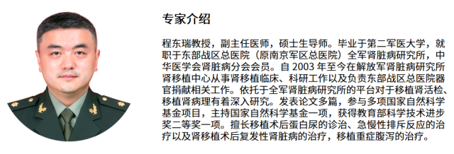
Q: Hello, Director Cheng! I recently saw that you participated in the European Annual Conference on Organ Transplantation (ESOT) in Copenhagen and shared the latest data on ddcfDNA in the study of antibody-mediated rejection (AMR) of transplanted kidneys with organ transplant experts around the world. Could you please explain to us why ddcfDNA is chosen as a marker for monitoring AMR in transplanted kidneys?
A: The full name of ddcfDNA is donor-derived cell-free DNA. The Chinese name is donor-derived cell-free DNA. It refers to the free DNA from the apoptosis or necrosis of donor cells in the circulating body fluids of patients after organ transplantation. It carries the health information of the donor tissue. Studies have shown that the concentration of ddcfDNA in kidney transplant recipients is closely related to transplanted kidney damage. Therefore, the health status of the transplanted kidney can be known by detecting ddcfDNA. The biological properties of cfDNA are relatively stable, easy to store and transport, and its half-life is short, which can reflect the patient's real-time health status. In addition, cfDNA is widely present in human blood and urine, and collection is convenient and non-invasive.
Q: Compared with our current clinical routine detection methods for rejection, such as serum creatinine, PRA, donor-specific antibodies (DSA) and renal biopsy, what are the clinical advantages of detecting ddcfDNA?
A: Serum creatinine index has hysteresis and low specificity. Renal tissue biopsy is invasive and carries risks of complications, and clinical judgment relies on the experience of pathologists. In addition, due to uneven pathological sampling and the early and late stages of disease progression, false negative results still exist. Data show that the false negative rate of biopsy is 20%-30%.
The positive predictive value (PPV) of DSA indicators for AMR is low. Our research data and internationally reported data show that the PPV of DSA is only 40%-45%. This shows that sometimes DSA is only in a free state and does not necessarily activate the damage pathways of vascular endothelial cells (complement-dependent pathway, direct interaction pathway with cell surface antigens and pathway of recruiting inflammatory cells). On the other hand, non-HLA antibodies (MICA, MICB, etc.) account for about 10%-40% of DSA. Currently, DSA mainly uses the Luminex-single antigen bead method platform to detect HLA-related antibodies, so it has a high false negative rate. Finally, for some patients with unclear donor HLA genotyping, it is difficult to determine whether DSA is present when their PRA is positive.
ddcfDNA is an early result of endothelial damage rather than a possible cause, and changes in AMR can be caused by either HLA or non-HLA antibody-mediated AMR. Our research data and internationally reported data both show that the PPV of ddcfDNA can reach 90%, which is much higher than the positive predictive value (~40%) of DSA alone in diagnosing AMR. In addition, ddcfDNA reflects the overall location of transplanted kidney damage, and there is no sampling heterogeneity.
Q: Is there any difference between ddcfDNA in detecting AMR and T cell-mediated rejection (TCMR)?
A: AMR and TCMR are different in the changes in plasma ddcfDNA concentration. The plasma ddcfDNA concentration is higher in AMR patients than in TCMR patients. According to Banff (2013) standards, AMR mainly causes damage to vascular endothelium, and the cfDNA produced after apoptosis of vascular endothelial cells directly enters the circulating blood. The main damage sites of TCMR are renal tubules and renal interstitium, and the apoptosis and necrosis of tubular epithelial cells into the blood must cross the vascular barrier. For rejection grade IB and above in TCMR (accompanied by endarteritis), research results have shown that plasma ddcfDNA is significantly increased. Further identification of rejection types requires the use of ddcfDNA information in multidimensional samples (urine and blood) to analyze different injury sites.
Q: How to use ddcfDNA to determine the risk of rejection? Can you share relevant clinical cases?
A: Different research results show that the cut-off value of plasma ddcfDNA concentration when detecting rejection is slightly different: A current study led by the University of Washington School of Medicine shows that when plasma ddcfDNA >1% is used as a threshold to diagnose rejection, the sensitivity can reach 81%, the specificity can reach 82%, and the AUC is 0.86. A study led by Cedars-Sinai Medical Center showed that the AUC for diagnosing rejection can reach 0.82. When plasma ddcfDNA ≥ 0.74%, the detection sensitivity can reach 100% and the specificity is 71.8%. Another study led by the University of Melbourne showed that the AUC for diagnosing rejection can reach 0.91. When plasma ddcfDNA>0.75%, the detection sensitivity is 85% and the specificity is 75%. Our research results on 88 kidney transplant patients in China showed that the AUC of using plasma ddcfDNA to detect rejection was 0.952. When plasma ddcfDNA was >0.96%, the detection sensitivity could reach 90% and the specificity could reach 96%.
We have encountered a female kidney transplant patient in clinical practice. After the transplant in March 2009, her creatinine was stable at 65 μmol. In March this year, her creatinine was 61.5 μmol, her urine protein was +, and there were no other clinical abnormalities. At this time, we performed a ddcfDNA test, and the result was that the concentration of plasma ddcfDNA was 3.72%, indicating a high risk of AMR. We further conducted a needle biopsy and showed that the patient had chronic transplanted kidney glomerulopathy. Compared with the 2010-01-02 section, the glomerular mesangial hyperplasia was accompanied by glomerulitis and glomerular waste. A small amount of peritubular capillaritis, mild tubulointerstitial lesions (15%), arterial hyaline degeneration and sclerosis. The final clinical judgment was subclinical rejection. This case may demonstrate that subclinical rejection can be detected early using plasma ddcfDNA.
Q: What is the main method for detecting ddcfDNA currently in the world?
A: The current mainstream technology for ddcfDNA detection in the world is the next generation high-throughput gene sequencing platform (NGS). NGS is the latest revolutionary technology in the field of life sciences and medical research, with a wide detection range, strong specificity and high sensitivity. It can detect thousands or even tens of thousands of SNP sites at one time, and the quantitative results are accurate. In addition, ddcfDNA detection methods also include qPCR, multiplex PCR, digital PCR, etc. Different quantitative methods of ddcfDNA have a certain impact on the detection results.
NGS technology can also be used to simultaneously diagnose BK virus risks, pathogenic microbial infections, delayed recovery of transplanted kidney function (DGF), etc., which can provide a more comprehensive indication of the causes of kidney transplant damage.
Q: What do you think is the future development direction of ddcfDNA testing in this field?
A: Currently, the single indicator of ddcfDNA concentration is mainly used to classify rejection and non-rejection reactions. In the future, the research model based on ddcfDNA and combined with other clinical indicators to predict the risk of rejection will be more scientific and accurate. The identification of rejection reaction types requires further use of ddcfDNA information in multidimensional samples to analyze different damage sites. In addition, other tissue-specific epigenetic features, such as cfDNA methylation, microRNA, nucleosome profiles, etc., have great potential in future research on different rejection damage sites. Finally, more research data is needed on the dynamic changes of ddcfDNA after treatment in rejection patients.
Thank you very much, Professor Cheng, for accepting our interview this time. Through this interview, we learned that donor-derived cell-free DNA plays a very important role in monitoring kidney transplant damage, and can detect kidney transplant rejection earlier with high specificity. We hope that there will be more research in this area in the future to help patients better monitor their kidney health and protect their lives and health.
References:
1. Cheng Dongrui et al., Research progress of donor-derived cell-free DNA in transplanted kidney injury [J]. Chinese Journal of Organ Transplantation (2019)
2. Lo YM, et al. Presence of donor specific DNA in plasma of kidney and liver transplant recipients[J]. Lancet (1998)
3. Roy D. Bloom et al. Cell Free DNA and Active Rejection in Kidney Allografts [J]. JASN (2017)
4. Whitlam JB, et al. Diagnostic application of kidney allograft-derived absolute cell-free DNA levels during transplant dysfunction [J]. AJT(2019)
5. Huang E, et al. Early clinical experience using donor-derived cell-free DNA to detect rejection in kidney transplant recipients [J]. AJT(2019)
Some original figures, videos and downloadable materials are provided in Chinese.