
AlloDx Viewpoint | Why do we need better Gd-IgA1 monoclonal antibodies?
IgA nephropathy (IgAN) is a common primary glomerular disease. Galactose-deficient IgA1 (Gd-IgA1) is considered to be the key initiating molecule in the multiple-hit pathogenesis of IgAN and one of the most potential serological markers. Over the past few years, Gd-IgA1 detection technology has been continuously iterated: from lectin methods to monoclonal antibody methods, from conventional ELISA to highly sensitive platforms such as time-resolved fluorescence. However, simply boosting the signal does not solve all problems. If the upstream antibody can only recognize a certain local sugar epitope in Gd-IgA1, then no matter how sensitive the detection platform is, it can only amplify part of the molecules that have been captured by the antibody. For patients who cannot be detected by existing methods, it is not necessarily because they do not have Gd-IgA1 in their bodies; it may also be that its main glycoform is not within the effective recognition range of current monoclonal antibodies.
Gd-IgA1 is not a structurally identical molecule
The IgA1 hinge region contains multiple potential O-glycosylation sites. During normal glycosylation, GalNAc will be further extended to form sugar chains containing galactose and other modifications; when galactosylation is insufficient, naked GalNAc and its surrounding peptides form complex sugar epitopes that can be recognized by antibodies. The problem is that galactose deletions can occur at different sites, with varying amounts, locations, and combinations of GalNAc exposures in different patients; adjacent glycans, Core 1 extension, and sialylation also alter the spatial accessibility of the epitope. Therefore, clinical Gd-IgA1 is more like a group of highly heterogeneous glycoform molecules than a fixed antigen.
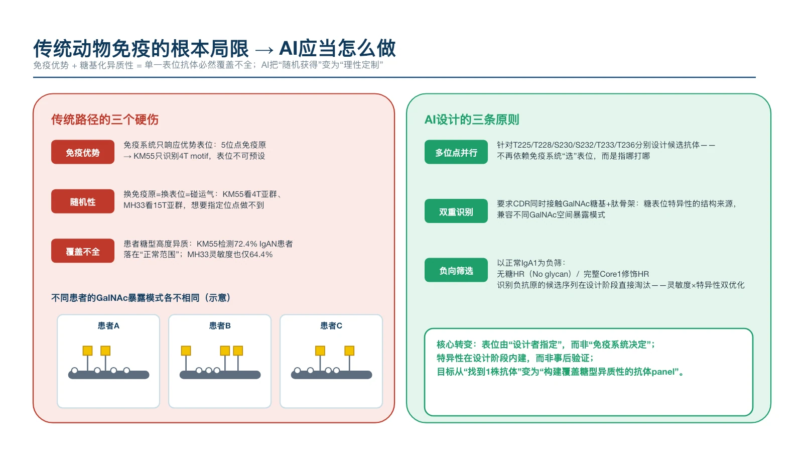
Figure 1 | Different patients may have different GalNAc exposure patterns; it is difficult for a single epitope antibody to completely cover glycoform heterogeneity.
The traditional development path has achieved today, but also limits the next step
Currently, Gd-IgA1 monoclonal antibodies mainly rely on in vitro synthesis of glycopeptides, animal immunization, and hybridoma screening. Researchers usually first synthesize a short peptide in the hinge region of IgA1, introduce GalNAc at one or more sites, couple it with a carrier protein, immunize mice or rats, and then screen candidate antibodies from a large number of hybridoma clones. This is a classic method that is executable at this stage and has been proven repeatedly. But it has four limitations that are difficult to completely eliminate from the process level:
01 Synthetic glycopeptides can only approximately simulate natural antigens
Intact IgA1 has a complex higher-order structure and multisite glycosylation. Preparing intact IgA1 with a clear, uniform and site-specific glycosylation state in vitro requires high technical difficulty, cost and batch-to-batch consistency control. Therefore, research and development often can only use short-chain synthetic glycopeptides, and it is difficult to completely reproduce the spatial conformation, adjacent sugar chain influence and hinge region accessibility in natural molecules.
02 There is an immune dominance bias in animal immunity
Even if an immunogen contains multiple GalNAc sites simultaneously, the immune system does not produce antibodies against each site equally. Dominant epitopes that are more spatially exposed and more likely to trigger immune responses tend to dominate the final clones obtained.
03 Hybridoma screening is inherently random
The traditional process involves first allowing animals to produce an antibody library and then looking for available antibodies from already formed B cell clones. Developers can choose the immunogen, but it is difficult to prescribe at what angle the antibody must approach the glycopeptide, or which sugar groups and amino acids must be contacted at the same time.
04 It is difficult for a single clone to cope with the heterogeneity of glycotypes in the population
When an antibody is biased towards a local glycoepitope, it may be under-responsive to patients carrying other glycosylation combinations. This epitope preference directly affects the population coverage of the test.
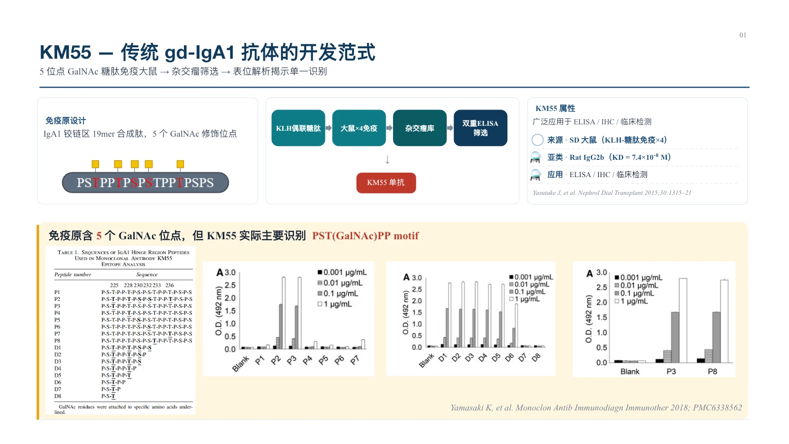
Figure 2 | The classic route represented by KM55: rats are immunized with multi-site GalNAc glycopeptides, but the final monoclonal antibody mainly recognizes the local motif near Thr225.
In 2015, the emergence of KM55 freed Gd-IgA1 detection from complete reliance on HAA lectin, improving the stability and standardization potential of the detection. However, subsequent epitope analysis found that although the immunogen contained multiple GalNAc modification sites, KM55 was mainly biased toward the PST(GalNAc)PP complex epitope near Thr225. MH33 reported in 2026 used Thr236 single-site GalNAc glycopeptide to immunize BALB/c mice to obtain antibodies biased towards the glycotope near Thr236. In clinical samples, the correlation between MH33 and KM55 detection results was low (IgAN group r=0.23, P=0.09), suggesting that the two antibodies may capture non-identical Gd-IgA1 molecule signals.
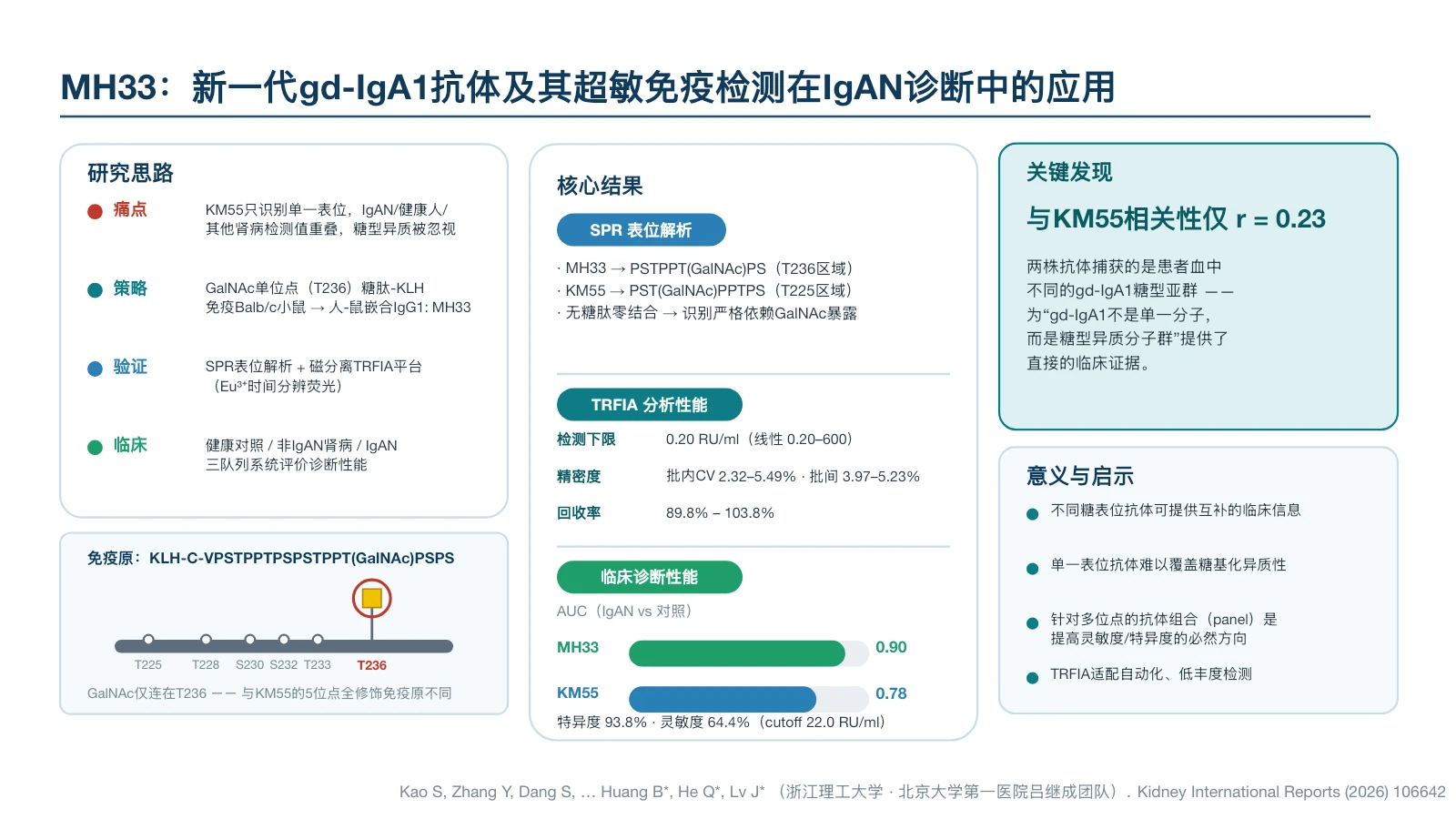
Figure 3 | MH33 and KM55 prefer different GalNAc glycoepitope, suggesting that epitope selection of monoclonal antibodies will change the detection signal of clinical samples.
Artificial intelligence is reshaping the path to antibody development
Currently, artificial intelligence and structural biology are offering a different path. The traditional method is to first let animals form an antibody library, and then search for the clone closest to the target from the library; AI-assisted design can first define the glycotope you want to recognize and the normal glycoform that needs to be rejected, and then generate and screen candidate sequences that better match these conditions.
1. Multi-site targeted design:Establish candidate structures for potential sites such as Thr225, Thr228, Ser230, Ser232, Thr233, Thr236, etc., without having to wait for the immune system to select epitopes for developers.
2. Dual recognition of glycosyl and peptide backbone:The candidate antibody's CDRs not only need to contact GalNAc, but also need to recognize its neighboring peptides to structurally establish site specificity.
3. Introducing negative screening during the design phase:Set the sugar-free hinge region, intact Core 1, and other normal IgA1 glycoforms as negative structures, and preferentially eliminate sequences where normal IgA1 cross-binding may occur.
4. Replace the "universal monoclonal antibody" assumption with combination coverage:Select complementary antibodies based on epitope and structural clustering to expand population glycoform coverage with dual or multi-antibody panels.
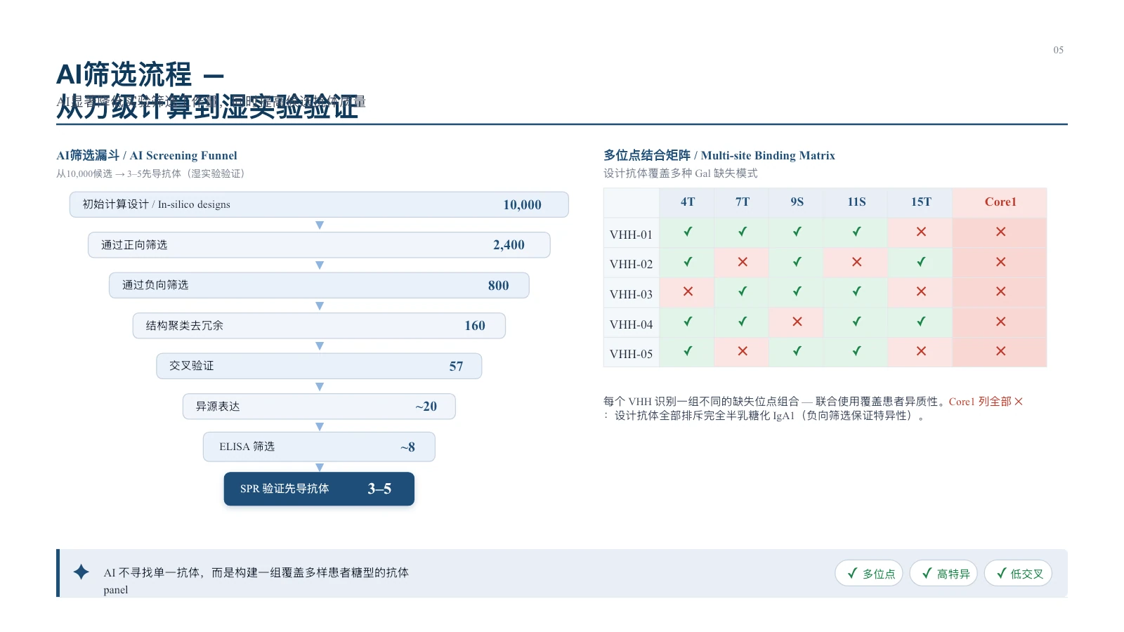
Figure 4 | The value of AI-assisted design lies in expanding the explorable sequence space and compressing a large number of computational candidates into a small number of experimental candidates through positive, negative and structural screening.
Of course, AI design cannot directly guarantee clinical performance based on calculation results. Candidate antibodies still need to undergo heterologous expression, synthetic glycopeptide binding, affinity analysis, normal IgA1 cross-reactivity, native Gd-IgA1 recognition and clinical cohort validation. The core value of AI is to allow wet experiments to start from a set of candidate sequences that are more consistent with the target structure, reducing the probability of relying entirely on random cloning.
What antibodies are needed for next-generation Gd-IgA1 testing?
A truly valuable candidate antibody should not just obtain a strong signal on a synthetic glycopeptide. It needs to go through a more stringent chain of evidence:
- The specified galactose-deficient site is clearly identified and its core epitope can be experimentally resolved;
- Recognizes GalNAc glycans simultaneously with adjacent peptide backbones rather than ubiquitin-binding glycans;
- Maintains low cross-reactivity to aglycopeptide, intact Core 1, and normal IgA1;
- Binding ability is retained in intact native IgA1 and patient samples;
- Have affinity, specificity, stability and manufacturability suitable for reagent development;
- Complementary with other epitope antibodies, demonstrating incremental value in combination in clinical cohorts.
|
Faced with the highly heterogeneous Gd-IgA1, instead of continuing to search for a universal antibody that "covers all glycoforms", a more feasible direction may be: directional design of multiple epitope antibodies, and then use clinical data to find the most complementary combination. |
Gd-IgA1 detection has completed an important leap from the lectin method to the monoclonal antibody method. The next upgrade may no longer only come from the signal detection platform, but will occur at the lower-level antibody design stage. When epitopes can be proactively defined by developers, when positive recognition and negative rejection can be considered simultaneously in the design stage, and when multiple antibodies can form complementary coverage around the glycoform heterogeneity of the population, only then can Gd-IgA1 detection break through the performance upper limit brought by a single epitope.
AlloDx is advancing Gd-IgA1 Research and validation of novel monoclonal antibodies in the hinge region.
Please stay tuned for subsequent technical progress.
References
[1] Yasutake J, et al. Novel lectin-independent approach to detect galactose-deficient IgA1 in IgA nephropathy. Nephrol Dial Transplant. 2015;30:1315–1321. DOI: 10.1093/ndt/gfv221.
[2] Yamasaki K, et al. Galactose-Deficient IgA1-Specific Antibody Recognizes GalNAc-Modified Unique Epitope on Hinge Region of IgA1. Monoclon Antib Immunodiagn Immunother. 2018;37:252–256. DOI: 10.1089/mab.2018.0041.
[3] Kao S, et al. Ultrasensitive Immunoassay Using a Novel Galactose-Deficient IgA1 Antibody and Its Clinical Application in the Diagnosis of IgAN. Kidney Int Rep. 2026;11:106642. DOI: 10.1016/j.ekir.2026.106642.
Statement:This information is used for technical and industry communication and does not constitute clinical diagnostic advice. The description of AI design in this article is the research and development direction and technology outlook; product performance and intended use should be subject to final experiments, clinical verification and registration approval.